top of page

VASCULAR & INTERNAL MEDICINE

Stormmy VoxelVu 3D Ventral

Stormmy VoxelVu 3D Dorsal

TURBO coronal MPR Still
Stormmy VoxelVu 3D Ventral
1/6
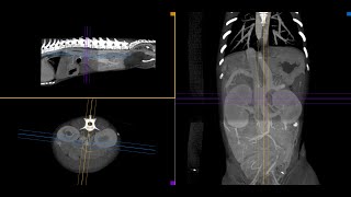
Experience unparalleled clarity and precision in veterinary CT imaging, with impeccable soft tissue detail and the industry's best contrast resolution, complemented by an unmatched spatial resolution of 300 to 600 microns.


VIMAGO™ GT30
VIMAGO™ GT30 Pico
For specialty or general practices that see a lot of vascular and internal medicine cases, we recommend either the Vimago™ GT30 or the Vimago™ GT30 Pico.
✔ Use HDVI CT with IV Contrast to scan to see what radiographs cannot show you.
✔ Accurately and rapidly diagnose shunts.
✔ PDA and PRAA in puppies are also diagnosed accurately and rapidly.
✔ Rapidly diagnose kidney function with IV contrast.
✔ Fully characterize masses and their possible association ureters, adrenals, and other anatomy.


“HDVI helps you make a definitive diagnosis. It helps in surgical planning, it helps immensely in dentistry and it is great for large dogs. Our practice uses the Vimago™ 3-5 times per day.”
Deepan Kishore, BVSC & AH, MS, ABVP Neel Veterinary Hospital

bottom of page